NIR-II fluorescence imaging
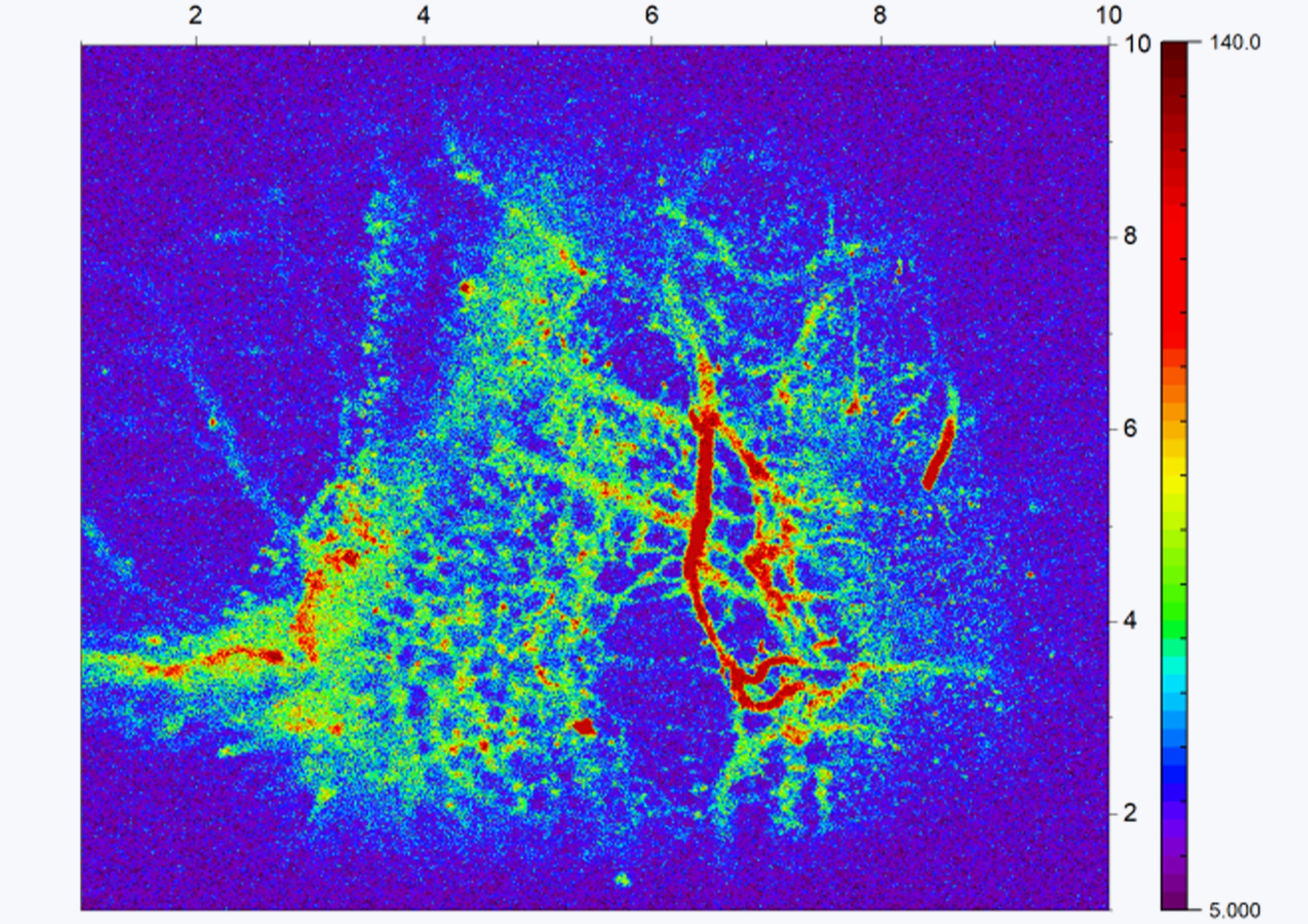
Lower tissue scattering and autofluorescence at 1000–1700 nm enable high-contrast imaging of deep tissue, vasculature and organs.

Connect confocal imaging, single-photon detection and time measurements around the fluorescence signal from your sample, for life science and materials research.
Twelve typical research directions across four fields: life-science imaging, biomedical probes, materials luminescence & spectroscopy, and quantum & low-dimensional materials. Each places different demands on excitation and emission bands, spatial and temporal resolution and signal level — use them to plan a Qumi configuration and related modules.
Lower tissue scattering and autofluorescence at 1000–1700 nm enable high-contrast imaging of deep tissue, vasculature and organs.
Confocal optical sectioning and 3D reconstruction for cellular structures, tissue sections and organoids.
Low-jitter superconducting detection provides lifetime contrast for microenvironment sensing, lifetime multiplexing and FRET.
NIR emission characterization and comparison of lanthanide nanoparticles, quantum dots, carbon nanotubes and small-molecule dyes.
Singlet-oxygen phosphorescence at 1270 nm is extremely weak; efficient, low-noise detection supports photodynamic therapy and oxidative-stress studies.
Track the distribution, targeting and metabolism of nanocarriers for theranostic research such as bone repair and oncology.
Photoluminescence (PL) imaging and spectral scanning to map defects, uniformity and emission efficiency.
Carrier lifetime and recombination dynamics for photovoltaic, light-emitting and photodetector materials.
NIR and SWIR emission, energy transfer and lifetime of rare-earth-doped materials.
Localization imaging, spectroscopy and antibunching of single quantum dots and color centers, combined with SNSPDs and timing modules.
Excitonic, defect-state and interlayer emission, with temperature-dependent spectroscopy in a cryostat.
NIR emission maps of silicon-photonic chips, lasers and detectors for performance evaluation and failure localization.
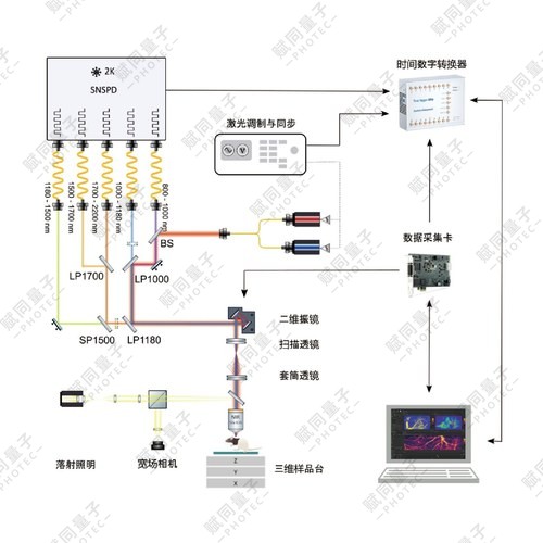
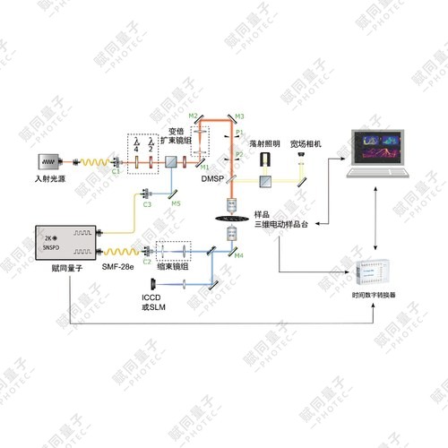
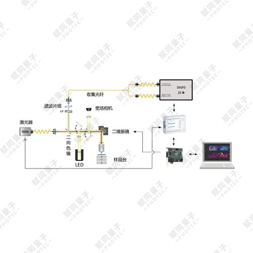
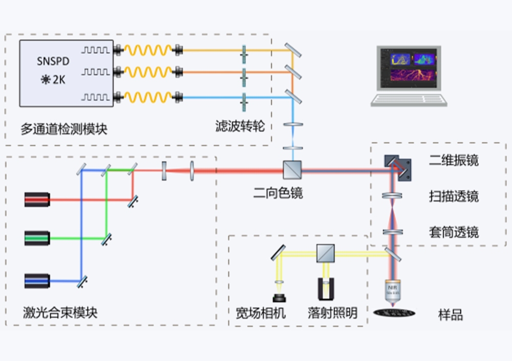
Near-infrared excitation is focused onto in-vivo, tissue-section or material samples.
Galvo scanning and confocal collection capture weak fluorescence point by point.
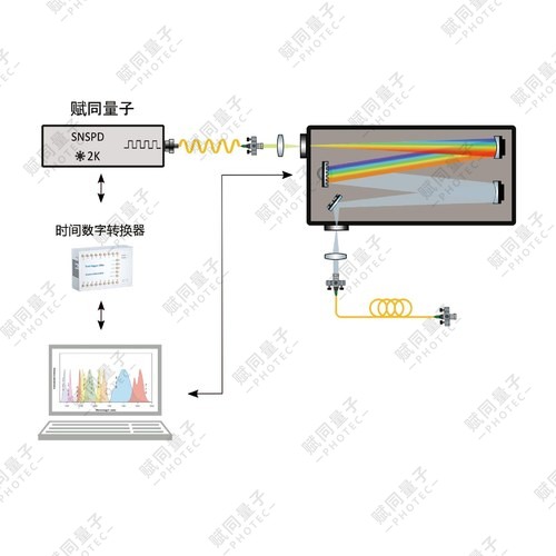
SNSPDs receive near-infrared photons with high efficiency and low dark counts.
Reconstruct 2D / 3D images, or run time-resolved analysis such as FLIM and TRPL.
An indicative experimental workflow. Modules and compatibility are confirmed for each configuration.
Explore single-photon confocal imaging and related measurement configurations.
Assess detection options against emission band and background signal.
Select time-correlated single-photon counting and accessories as required.
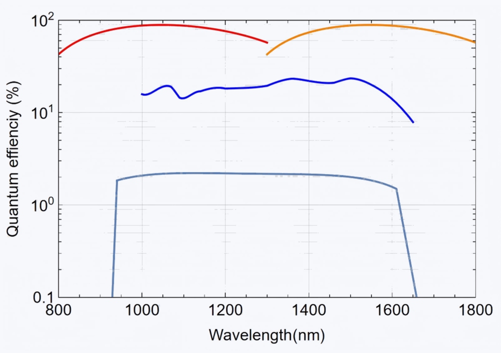
Qumi-2000 detects fluorescence with superconducting nanowire single-photon detectors, offering higher detection efficiency (peak quantum efficiency >90% at 1550 nm), one to two orders of magnitude lower noise and lower timing jitter.
Share your sample, measurement goals and existing equipment so we can discuss a suitable configuration.