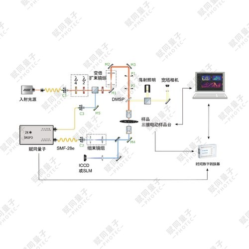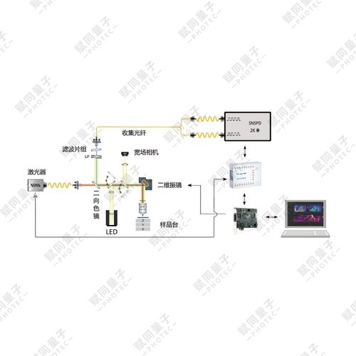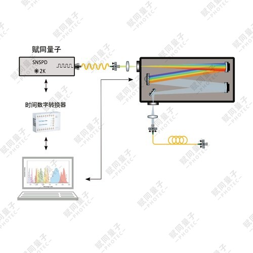
The system uses a superconducting nanowire single-photon detector (SNSPD) as the core receiver. Superconducting single-photon detection delivers high efficiency and low noise across 800–2200 nm, overcoming the weak long-wavelength response and high noise of PMT and InGaAs detectors.
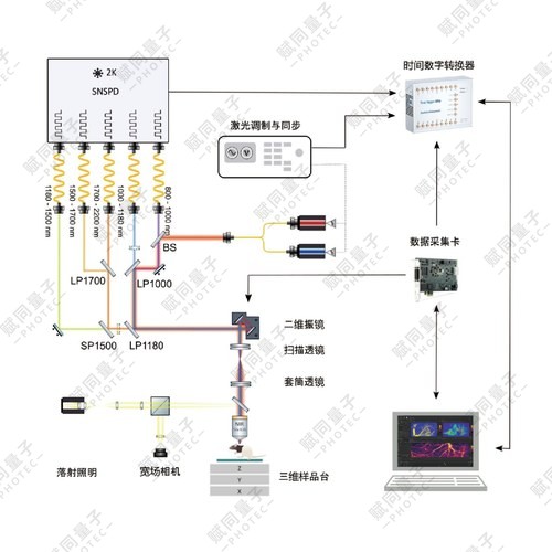
Product details
Qumi-Micro · Reflective single-photon microscopy
A 5-channel, 800–2200 nm single-photon microscopy platform for intensity, lifetime and time-gated imaging.
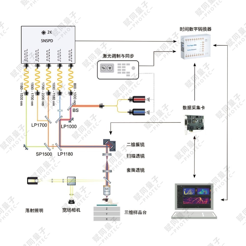
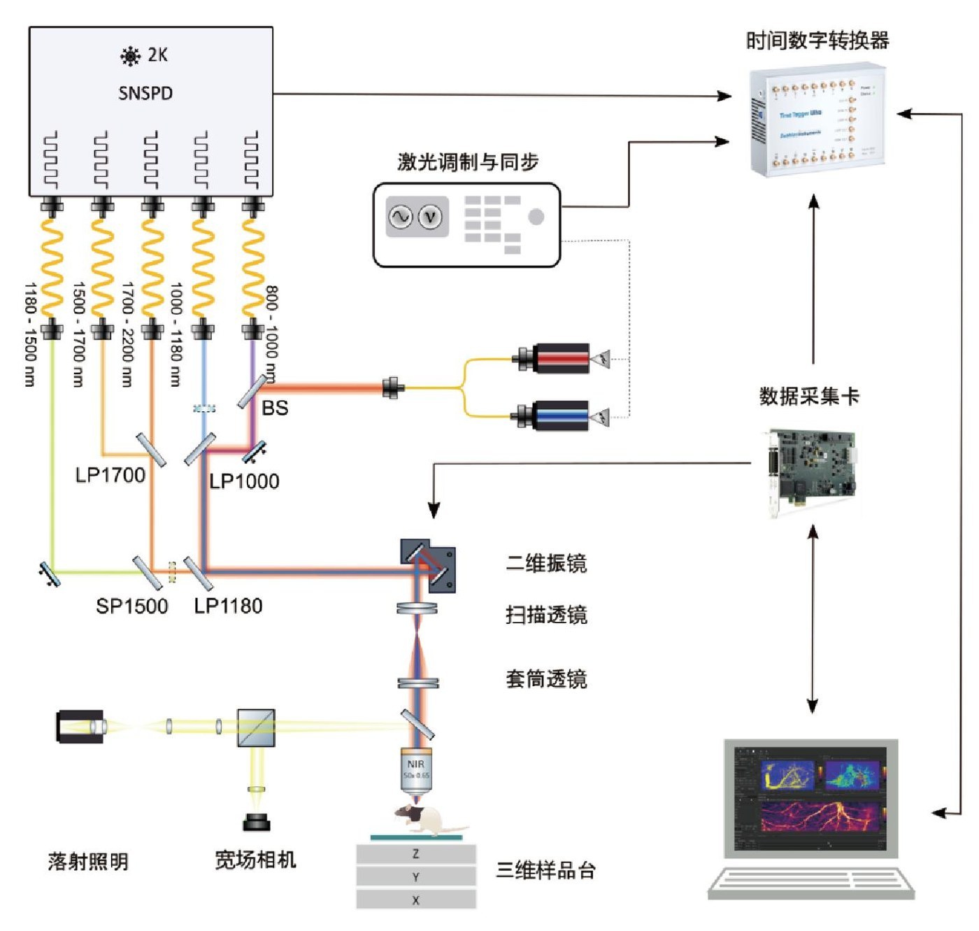
Qumi-Micro reflective microscopy combines SNSPDs with scanning, microscope optics, a motorized XYZ stage and data-acquisition software for near-infrared weak-fluorescence experiments. It supports single/multicolour 2D and 3D imaging, time series, ROI scans, large-field stitching, FLIM, time gating and photon statistics.
800–2200 nmDetection wavelength range
>80%Detection efficiency
5 channelsExpandable detection channels
~50–100 psTiming resolution
Core capabilities
- 808 nm and 980 nm CW/pulse-modulated excitation, expandable to other wavelengths.
- 1 fps at 128×128; up to 4096×4096 pixels and 0.1 ms–1 s pixel dwell time.
- 5 SNSPD channels with single-mode/multimode fiber coupling and channel expansion.
- Integrates FLIM, time gating, photon statistics and multidimensional imaging.
System schematic
Technical specifications
| Module | Parameter | Standard configuration | Expansion / options |
|---|---|---|---|
| Excitation source | Lasers | 808 nm, 980 nm (CW / pulse-modulated) | Other excitation wavelengths supported |
| Scanning module | Scan speed | 1 fps @128×128 | Customisable / upgradable |
| Scanning module | Pixel dwell time | 0.1 ms–1 s | |
| Scanning module | Image resolution | Up to 4096×4096 | |
| Scanning module | Optical resolution | ~1 μm @1550 nm SMF | |
| Scanning module | Timing resolution | ~50–100 ps | |
| Detection module | Detector | Superconducting nanowire single-photon detector (SNSPD) | |
| Detection module | Detection wavelength range | 800–2200 nm | Optional extension to 400–1350 nm (visible and near-infrared) |
| Detection module | Detection efficiency | >80% | |
| Detection module | Dark noise | <100 Hz (single-mode); <3000 Hz (multimode) | |
| Detection module | Channels | 5 | Additional detection channels available |
| Detection module | Coupling | Single-mode / multimode fiber | |
| Microscope body | Objective | NIR infinity-corrected plan apochromat; 400–1800 nm, 20×, NA=0.4 | Other objective magnifications available |
| Microscope body | Wide-field illumination | Visible epi-illumination | Optional fiber mercury lamp, LED, etc. |
| Microscope body | Camera | Visible-to-NIR imaging, 400–1100 nm | Optional SWIR camera (400–1700 nm) |
| XYZ sample stage | XY axes | Motorised XY, 100 mm travel; repeatability ±100 nm; minimum step 10 nm | Customisable / upgradable |
| XYZ sample stage | Z axis | Motorised Z, 50 mm travel; repeatability ±500 nm; minimum step 200 nm | Customisable / upgradable |
| XYZ sample stage | Sample platform | 185×185 mm; maximum load 3 kg | Customisable / upgradable |
| Acquisition and software | Acquisition hardware | DAQ: 4 high-speed analog output/input channels and 4 counters; TDC | PMT detection compatible |
| Acquisition and software | Computer | Multicore high-speed processor, 32 GB RAM, 500 GB SSD | |
| Acquisition and software | Imaging modes | Single/multicolour XY, XYZ, XYT, XYZT; full-field / ROI scans and large-field stitching; FLIM, time gating and photon statistics | |
| Other options | NIR-II wide-field imaging | Optional on request | SWIR camera and high-power excitation source |
| Other options | Spectral detection | Optional on request | Spectrometer for fluorescence spectroscopy |
Applications
Brain vasculature and in-vivo fluorescenceNIR-II fluorescence-intensity scans and multiband imaging.
Fluorescent probe evaluationIntensity and lifetime information characterize sample emission.
Fluorescence lifetime imagingFLIM and time gating investigate spatiotemporal emission.
Weak-light material characterizationMicro-region luminescence and photon statistics at low photon flux.
Imaging results
Brain vasculature fluorescence intensity imaging
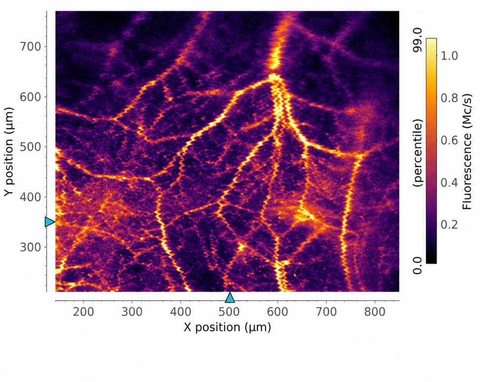
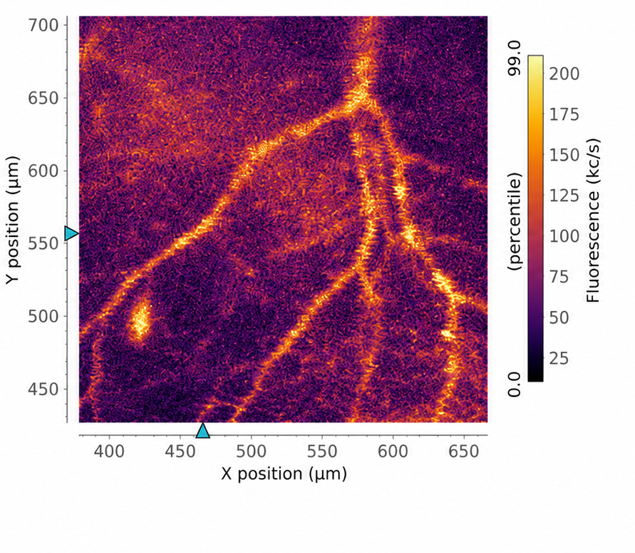
Mouse brain vasculature after injection of rare-earth nanoparticles. Left: 1500–1700 nm; right: 1700–2200 nm.
Fluorescence intensity and lifetime comparison
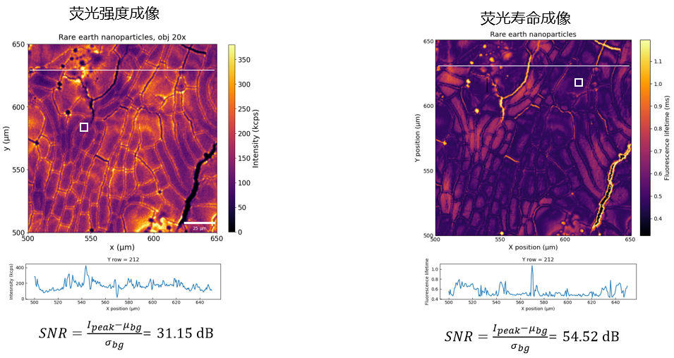
Fluorescence intensity (left) and lifetime (right) imaging of the same sample. SNR is 31.15 dB and 54.52 dB respectively in this example, an increase of about 23.37 dB with lifetime imaging.
Configuration and options
Detector and objective wavelength ranges are listed separately. Effective imaging range depends on the complete optical and detection configuration. Single-mode and multimode coupling have different dark-noise specifications. Confirm optional and expanded configurations for the experiment. Results illustrate the stated samples and experimental conditions.