To counter the coupled intrinsic noise sources in ultra-low-light microscopy, the platform suppresses non-target noise at the source along the whole optical chain — laser ASE, Raman/Rayleigh scattering in optics and substrate autofluorescence — achieving an imaging SNR above 50 dB in hardware without relying on frame accumulation or algorithmic denoising.
Qumi-Micro ultra-low-light microscopy system
Product details
Qumi · Single-photon imaging and spectroscopy
Optical noise suppression and photon time tagging for extremely low-flux microscopy.
The platform suppresses coupled optical noise sources throughout the system, including laser amplified spontaneous emission, Raman/Rayleigh scattering and substrate autofluorescence. This reduces reliance on frame accumulation and software denoising, achieving weak-light imaging SNR above 50 dB in the hardware optical chain.
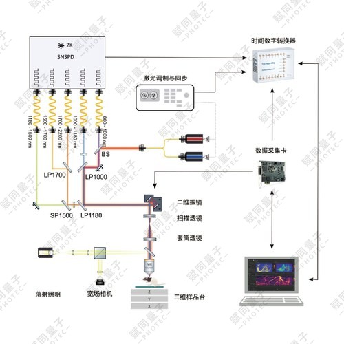
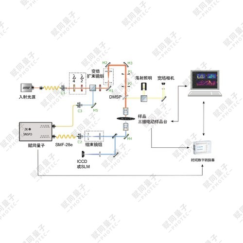
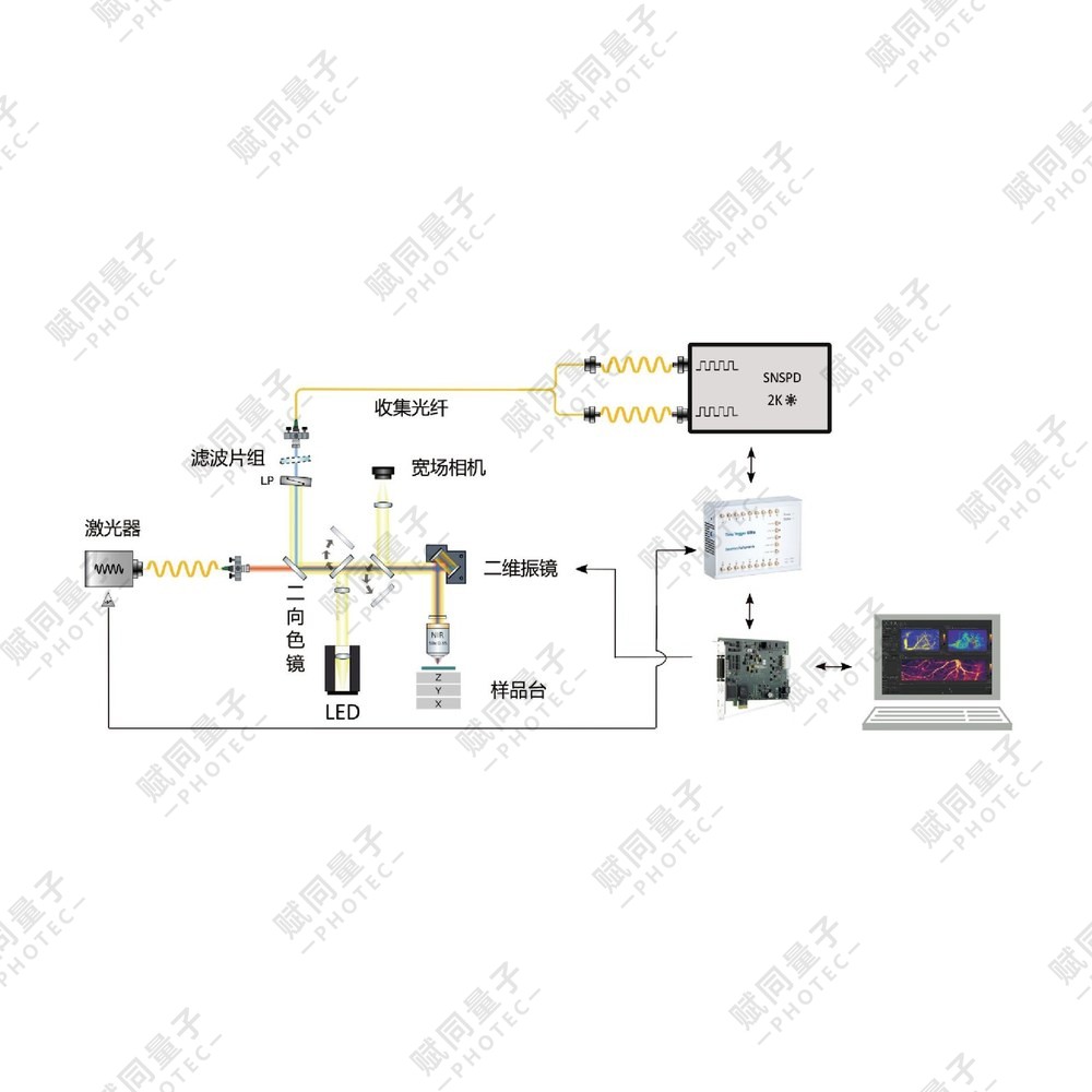
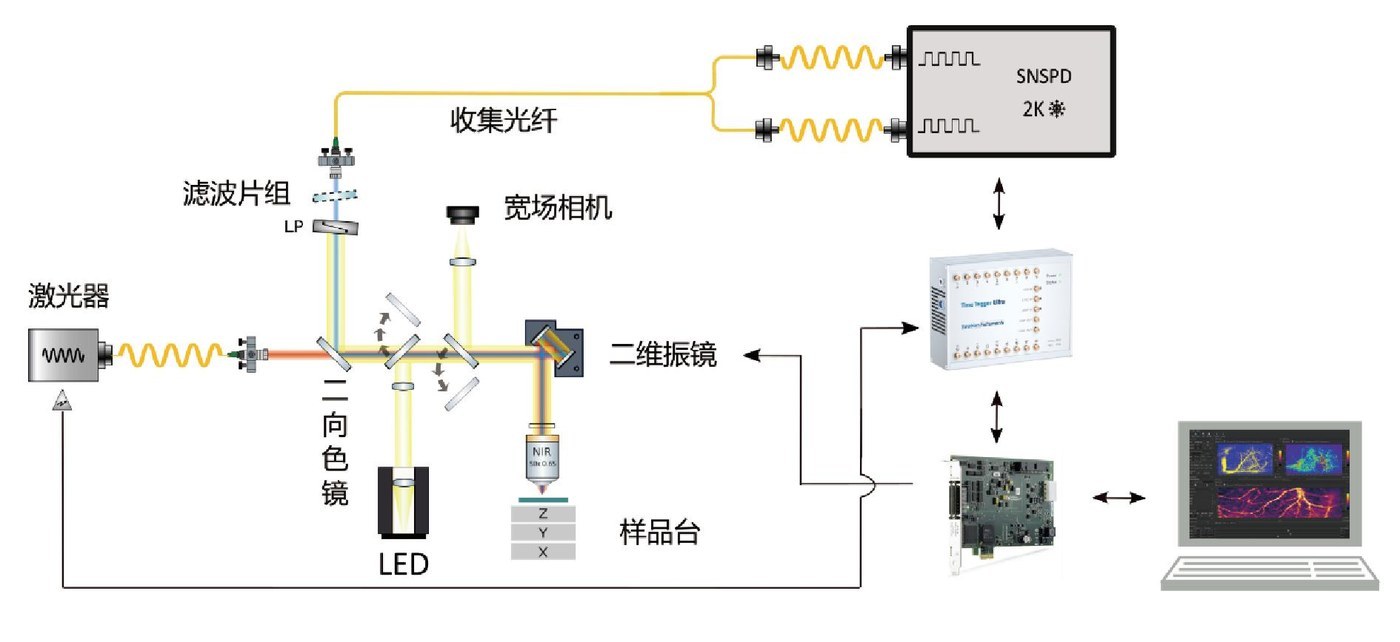
A modular optical architecture supports custom excitation and detection paths. Precision XYZ nanopositioning combines with wide-field preview, confocal sectioning, dynamic time-series imaging and native photon time tagging. Images and photon metadata support common research analysis tools.
The system addresses single-molecule fluorescence, 2D defects, crystal color centers, semiconductor quantum dots and rare-earth emitters. Low excitation flux supports imaging with reduced photobleaching and phototoxicity.
400–1800 nmWavelength band
10 nmMinimum XY closed-loop step
Picosecond classTime resolution
Photon time taggingData acquisition
Core capabilities
- Full-path optical noise suppression.
- Modular excitation and detection paths can be customized.
- Integrates wide-field preview, reflection confocal imaging, XYZ sectioning and XYT imaging.
- Saves multiple image formats and complete photon metadata.
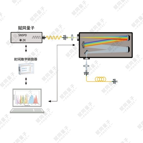
System schematic
Technical specifications
| Parameter | Configuration |
|---|---|
| Wavelength band | 400–1800 nm |
| Objectives | General-purpose: 5×, 10×, 20× long-working-distance achromats; high magnification: 50× / NA 0.65 |
| Precision XY scanning stage | Travel: 100×100 mm; minimum closed-loop step: 10 nm |
| Precision Z coarse stage | Travel: 30 mm; minimum closed-loop step: 0.1 μm |
| Timing control and acquisition | Stage TTL pulses at equal displacement intervals; picosecond-class TDC acquisition |
| Imaging modes | Wide-field preview, reflection confocal, XYZ sectioning and XYT dynamic time series |
| Data | Multiple image formats and complete metadata, compatible with common analysis tools |
Applications
Single-molecule fluorescenceLocalisation, imaging, blinking dynamics and transition-dipole orientation.
2D material defect statesEmitter screening and density statistics, saturation characteristics and single-photon purity.
Crystal color centersDensity mapping, antibunching, spin-related fluorescence changes and saturation curves.
Semiconductor quantum dotsFluorescence switching statistics, spectral/intensity inhomogeneous broadening, exciton fine structure and quantum yield.
Rare-earth emittersUpconversion/downconversion, nonlinear intensity dependence and long-lifetime time-resolved dynamics.
Configuration and options
Optical, detection and excitation modules can be configured for the sample emission and experiment.