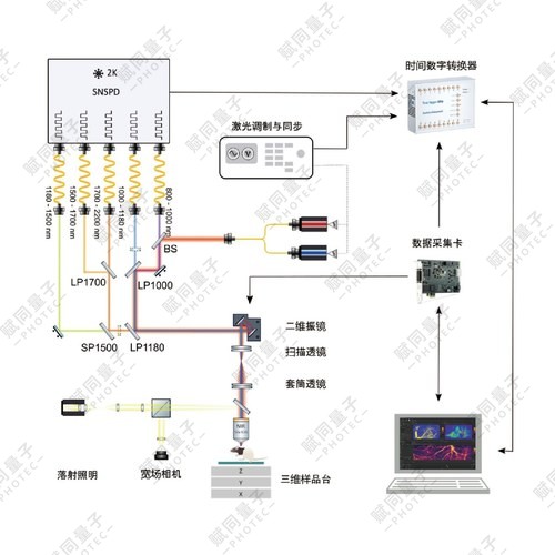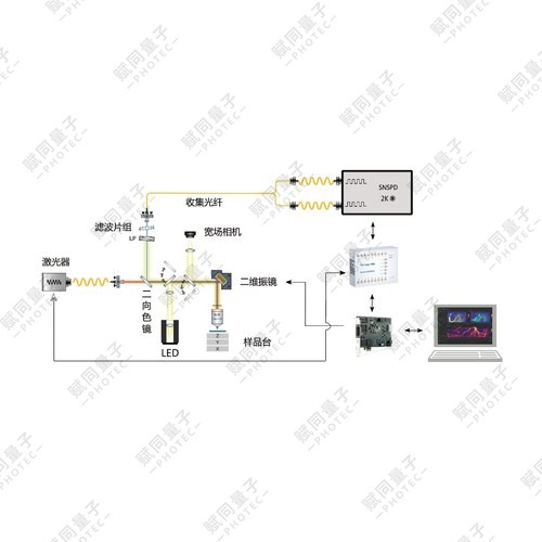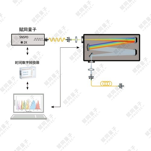
Conventional reflective confocal microscopes collect back-scattered signal, so much of the information carried by transmitted light from waveguides, thin films and transparent biological samples is lost; under weak light the transmitted photon flux is easily masked by stray reflected light.
Qumi-Micro transmission microscopy system
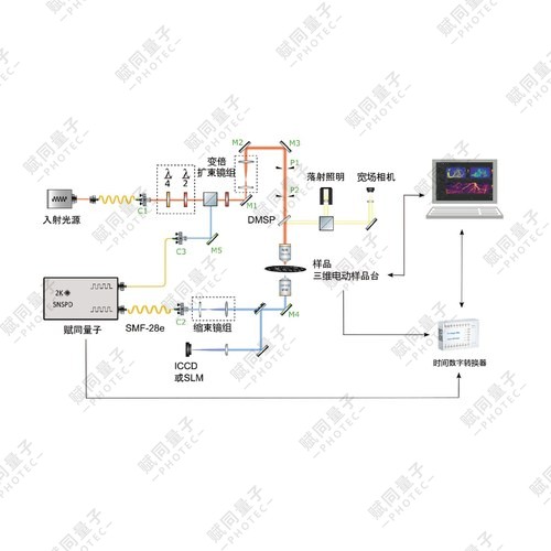
Product details
Qumi · Single-photon imaging and spectroscopy
Separate illumination and transmission paths for weak-light microscopy of transparent samples.
Conventional reflective confocal microscopy collects backscattered light, losing important transmitted information from waveguides, films and transparent biological samples. At low flux, transmitted signals can be obscured by stray light in the reflection path.
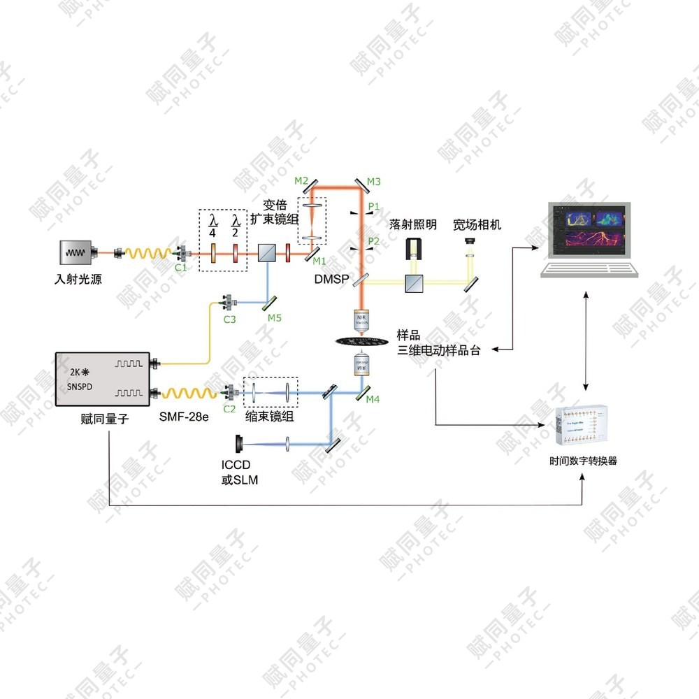
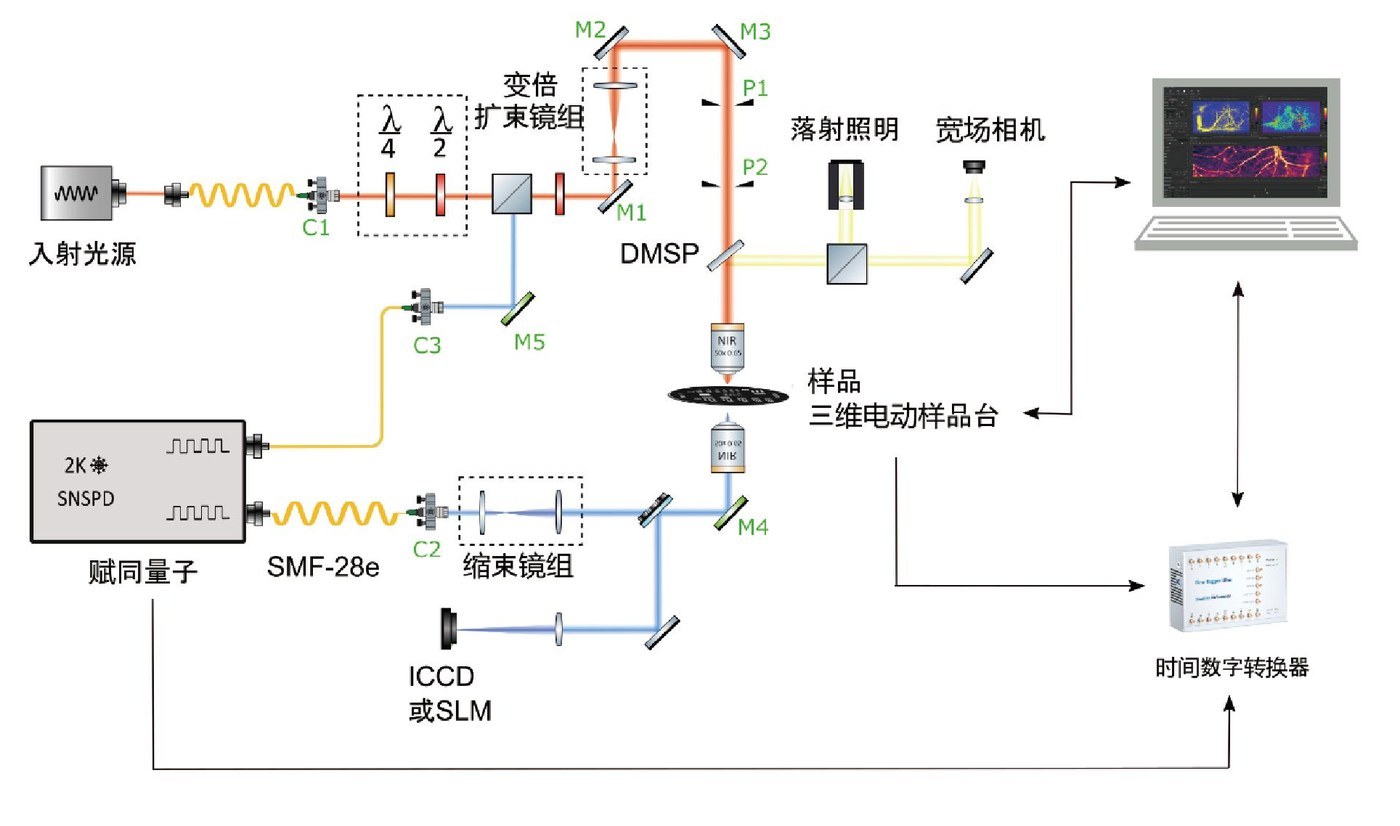
A dual-objective architecture separates illumination and collection: the upper objective focuses the excitation, while the lower high-NA objective collects transmission. The platform integrates wide-field preview, transmission and reflection confocal microscopy, XYZ sectioning and XYT time-series imaging. It supports label-free 3D reconstruction of transparent samples for quantum optics, integrated photonics, 2D materials and near-infrared biological imaging.
1100–1800 nmWavelength band
~1 μmLateral resolution at 1550 nm
10 nmMinimum XY closed-loop step
2 nmMinimum piezo closed-loop step
Core capabilities
- Dual objectives separate illumination from transmission collection.
- Integrates wide-field preview, transmission/reflection confocal imaging, XYZ sectioning and XYT imaging.
- Motorised stages combine with piezo fine positioning and three-point leveling.
- Stage TTL triggering synchronizes with TDC timing acquisition.
System schematic
Technical specifications
| Parameter | Configuration |
|---|---|
| Wavelength and resolution | 1100–1800 nm; lateral: ~1 μm @1550 nm; axial: ~5 μm @1550 nm |
| Optical coupling and alignment | Single-mode fiber coupling efficiency: >80% |
| Objectives | Dual-objective coupling efficiency: ≥90%; general-purpose: 5×, 10×, 20× long-working-distance achromats; high magnification: 50× / NA 0.65 |
| Precision XY scanning stage | Travel: 100×100 mm; minimum closed-loop step: 10 nm |
| Precision Z coarse stage | Travel: 30 mm; minimum closed-loop step: 0.1 μm |
| Piezo nanopositioning stage | Fine travel: 500 μm; minimum closed-loop step: 2 nm |
| Sample leveling | Three-point nanoscale leveling; pitch/tilt angular resolution: 0.00133° |
| Mechanical stability | Over 12 h, transmission efficiency decrease ≤5% |
| Timing control and acquisition | Stage TTL pulses at equal displacement intervals; picosecond-class TDC; line-by-line fly scanning with low serial latency and no pixel misalignment |
| Imaging modes | Wide-field preview, transmission confocal, reflection confocal, XYZ sectioning and XYT dynamic time series |
Applications
Quantum optics and integrated photonicsNear-field characterization of metasurfaces, waveguide mode imaging, single-photon devices and SNSPD-based weak-signal detection.
Optoelectronic materials and micro/nanostructuresNondestructive transmission characterization of films, photonic crystals and 2D materials; defect sectioning and 3D morphology of semiconductor structures.
Bioimaging and life sciencesIn-vivo imaging in the 1100–1800 nm biological window, label-free 3D sectioning of transparent tissues and time-series monitoring of cellular processes.
Configuration and options
Wavelength band, objectives, stage travel and acquisition modules can be configured for the experiment.