
01 Introduction
This collaboration using the Qumi platform for extremely weak light detection relies on Photec superconducting nanowire single-photon detectors (SNSPDs) for deep-tissue confocal imaging and weak fluorescence collection in the long-wavelength NIR-II window. The work was published in Advanced Imaging.
With reduced tissue scattering and a greater proportion of ballistic photons, the long-wavelength NIR-II window is an important direction for deep in-vivo optical imaging. However, water absorption is significant in this range, and biocompatible organic fluorescent probes emit very little light in their long-wavelength tails. Conventional free-space confocal optics are also susceptible to mechanical drift and alignment errors. Unstable excitation–detection conjugation compromises background rejection when photons are scarce, limiting contrast and resolution at depth.
Recently, teams led by Professor Jun Qian at Zhejiang University and Researcher Jie Liu at Nanjing Tech University introduced fiber wavelength-division multiplexing (WDM) into a confocal microscope. Combined with sensitive SNSPD detection, the architecture enabled high-resolution imaging of cerebral blood vessels in living mice at depths of up to 900 μm, using the weak emission tail of organic nanoprobes under photon-limited conditions.
02 Fiber WDM confocal architecture: maintaining optical conjugation in weak light
The central challenge in long-wavelength NIR-II deep-tissue imaging is that fluorescence is already extremely weak. Conventional free-space confocal systems use dichroic mirrors to separate excitation and detection. Vibration and optical misalignment can degrade coaxial conjugation, reducing the confocal pinhole’s background filtering efficiency and allowing noise to overwhelm the signal.
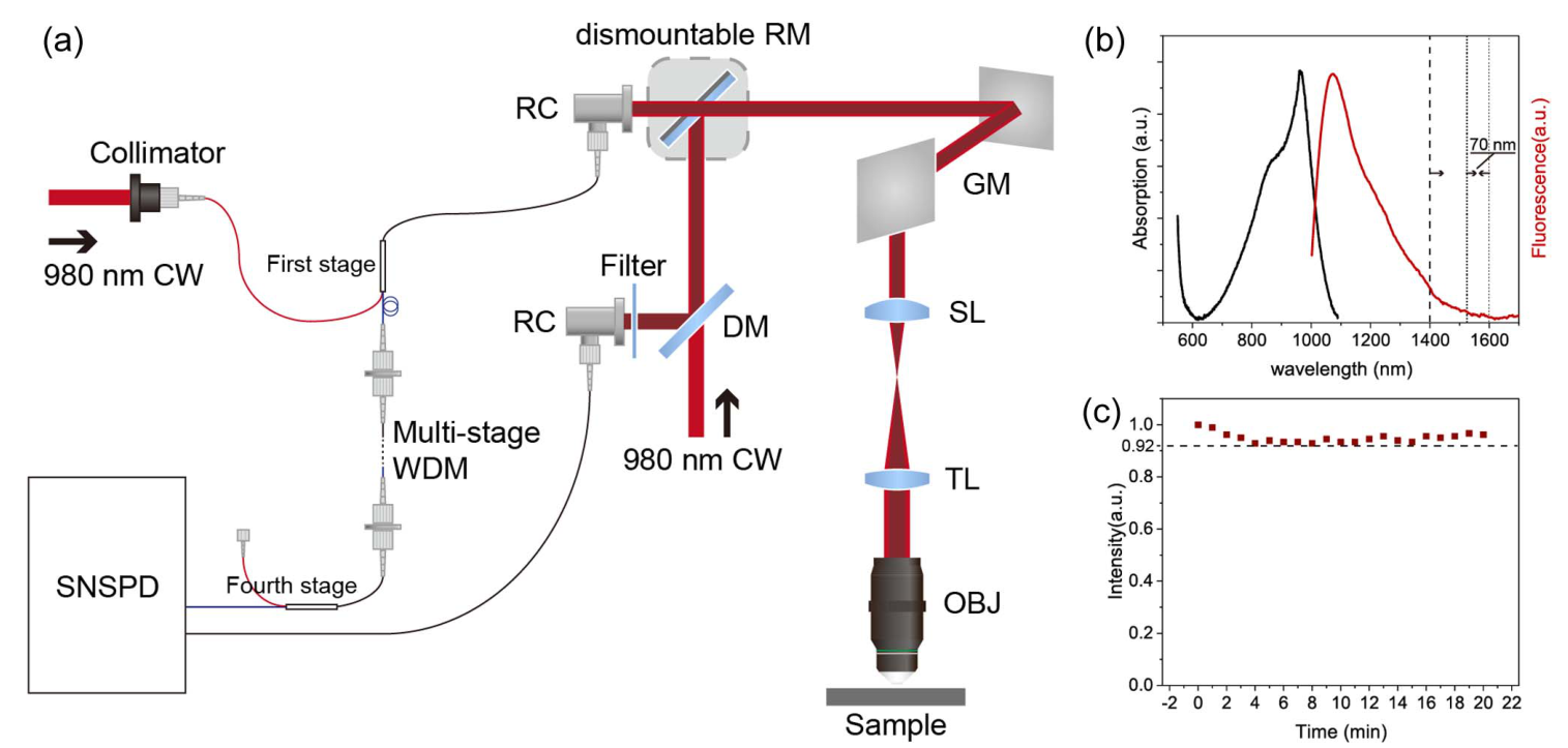
The researchers adapted WDM technology from fiber-optic communications, using four cascaded 980/1550 nm WDM modules. Excitation light and fluorescence travel coaxially through the same fiber and share the same collimation unit, establishing intrinsic excitation–detection conjugation at the system level.
Separating wavelengths within the fiber avoids conjugation degradation caused by free-space alignment errors and maintains stable confocal filtering without repeated alignment. Background rejection is preserved even when very few photons are available in long-wavelength deep-tissue imaging, providing a robust foundation for weak-fluorescence measurements.
03 Using a weak fluorescence tail to image 900 μm deep in vivo
The study used biocompatible L1057 organic nanoparticles with an emission peak at 1057 nm. Imaging detection instead used the long-wavelength tail at 1530–1600 nm, which accounts for only about 2% of total fluorescence. Strong water absorption in biological tissue further reduces this already limited photon budget.
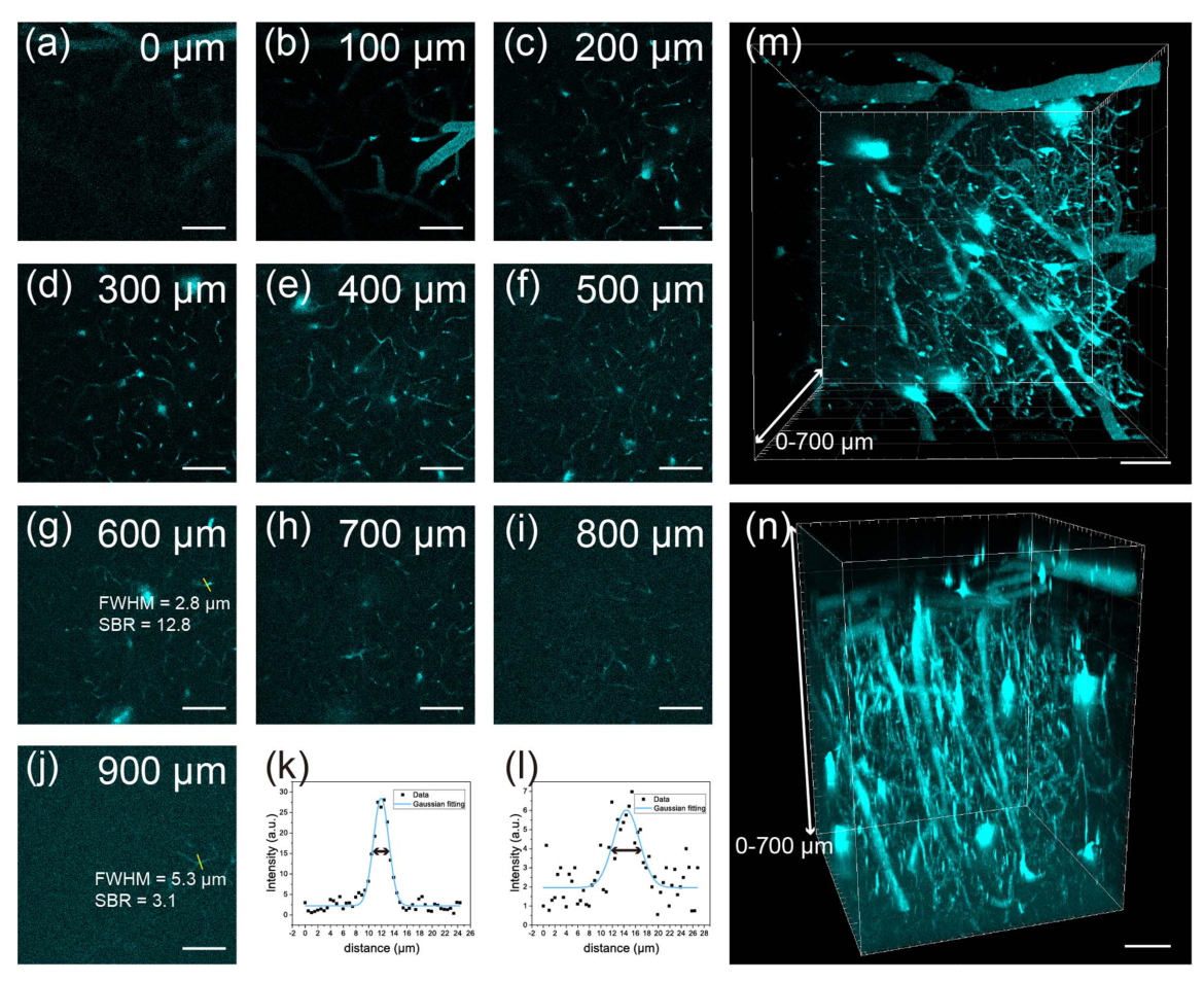
In experiments on cerebral blood vessels in living mice, the WDM confocal system captured continuous vascular structures from the cortical surface to a depth of 900 μm. Clear three-dimensional reconstruction was possible over 0–700 μm. Under the same probe and excitation conditions, the conventional free-space confocal system reached approximately 600 μm.
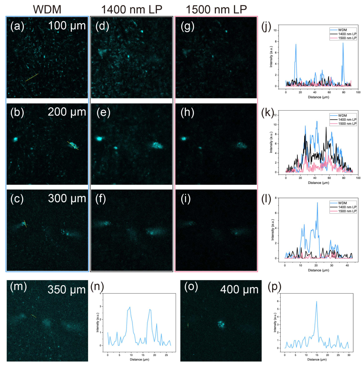
At 600 μm, the measured lateral full width at half maximum (FWHM) of a vessel profile was 2.8 μm, with a signal-to-background ratio of 12.8. At the maximum depth of 900 μm, a microvessel profile retained a FWHM of 5.3 μm and a signal-to-background ratio of 3.1, allowing fine vascular structures to remain distinguishable. In highly scattering ex-vivo liver tissue, same-field comparisons showed that the WDM approach outperformed conventional long-pass-filter configurations at depths of 100–300 μm and extended imaging to 400 μm, demonstrating its advantages in strongly scattering tissue.
04 Qumi series: sensitive detection for deep-tissue NIR-II imaging
Stable imaging also depends on sensitive single-photon detection. In this experiment, long-wavelength fluorescence was coupled directly from the final WDM module’s 1550 nm port into a Photec SNSPD for precise photon-counting acquisition.
Around the 1550 nm NIR-II window, Photec SNSPDs combine high detection efficiency with very low dark count rates. Their compatibility with the WDM system’s small-core single-mode fiber helps preserve scarce fluorescence photons. This reduces the limitations associated with the dark counts and sensitivity of conventional InGaAs detectors and photomultiplier tubes, allowing weak long-wavelength emission tails to produce stable, interpretable imaging signals.
Based on Photec’s superconducting single-photon detection technology, the Qumi series supports NIR-II biological imaging and materials characterization through:
- Confocal fluorescence imaging and deep-tissue observation in the NIR-IIb/NIR-IIc bands
- Weak-light steady-state spectroscopy and time-resolved fluorescence lifetime measurements
- Customized experimental support, including multichannel photon counting and ratiometric imaging
The Shanghai Qumi Demo Laboratory is open for sample measurements and preliminary feasibility studies. Researchers working with rare-earth nanocrystals, quantum dots, new long-wavelength organic dyes and other fluorescent probes are welcome to explore applications at the intersection of biomedical and materials science.
Paper DOI: 10.3788/AI.2026.10003